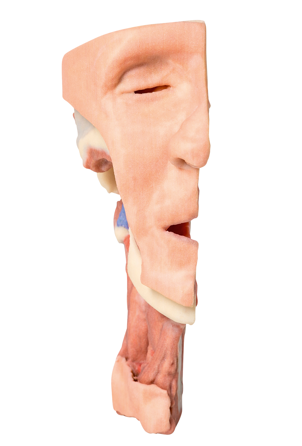
MP1665 - Deep face/Infratemporal fossa
MP1665 - Deep face/Infratemporal fossa
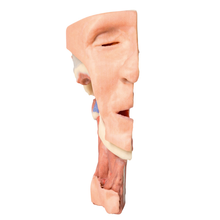
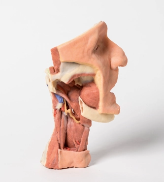
In this specimen the ramus, coronoid process and head of the mandible have been removed to expose the deep part of the infratemporal fossa. The pterygoid muscles have been removed to expose the lateral pteygoid plate and posterior surface of the maxilla. The buccinator has been retianed and can be seen originating from the outer aspect of the maxilla, the pterygomandibular raphe and the outer aspect of the mandible (which edentolous in this specimen).The superior constrictor arises from the posterior aspect of the pterygomandibular raphe.
The internal laryngeal nerve has been preserved. Muscles in the neck that are identifiable include mylohyoid, the strap muscles and the inferior constrictor.
The styloid muscles can be seen descending from the process to their insertions (not shown). The internal carotid artery can be seen deep to the styloid process which gives origin to stylohyoid, styloglossus and stylopharyngeus.
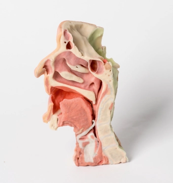
On the medial aspect of the sagittal surface the features of the lateral wall of the nasal cavity (superior, middle and inferior conchae and sphenoethmoidal recess, superior meatus, middle meatus and inferior meatus), the nasopharynx, the opening of the auditory tube, the hard palate, soft palate, oropharynx, laryngopharynx, hyoid bone (white) and laryngeal carticlages (blue/grey). The muscles of the tongue are discernible. The parts of the larynx and the pharynx are clerly seen. The verterbral bodies of C2-C5 as well as the arch of C1(atlas) and the dens of C2 or axis are clearly seen in the mid sagittal cut.
- Quantitative unit
- ks
MP1665 - Deep face/Infratemporal fossa
