MP1600 - Circle of Willis
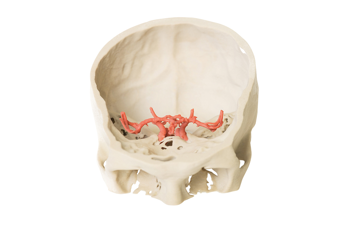
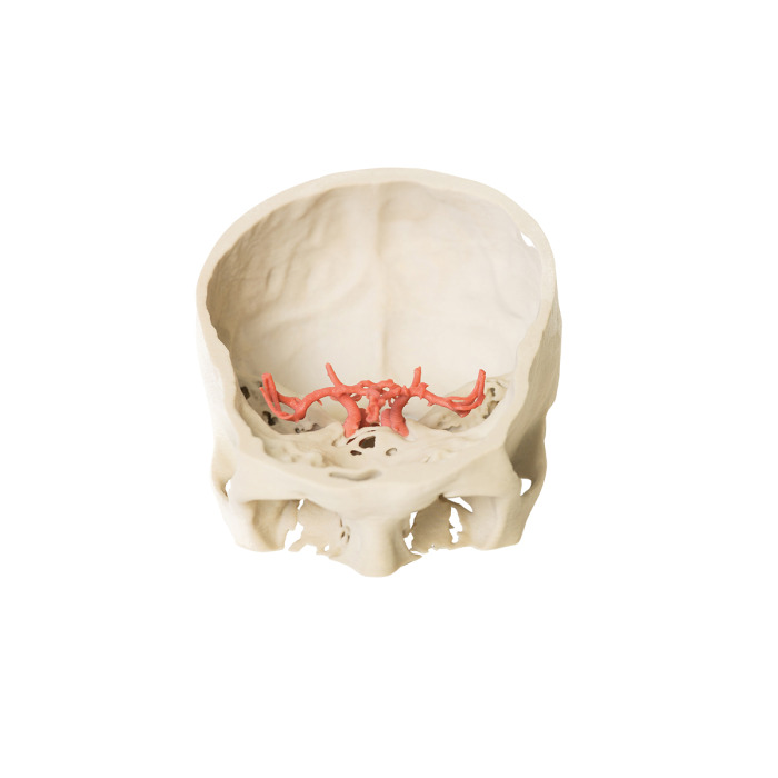
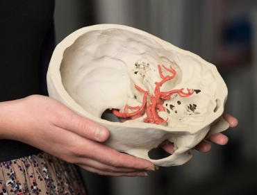
This model demonstrates the intracranial arteries that supply the brain. The model was created by careful segmentation of angiographic data. There would be no physical manner in which to dissect these arteries in situ as displayed. The model shows the paired vertebral arteries entering the cranial cavity through the foramen magnum and uniting to form the basilar artery. The basilar can be seen dividing into their terminal posterior cerebral arteries. The superior cerebellar arteries arise just proximal to this termination.
The internal carotid arteries (ICAs) can be traced from the point where they enter the skull base at the carotid canal on the temporal bone and travel medially and anteriorly within the canal in the petrous part of the temporal bone to emerge through the upper opening of the foramen lacerum. It is here that each ICA lies within the cavernous sinus (not shown). The S-shaped carotid siphon on both left and right sides are most beautifully demonstrated lateral to the sella turcica . They then pass medial to the anterior clinoid processes. They then divide into anterior and middle cerebral arteries. The paired posterior communicating arteries are clearly visible connecting the posterior cerebral and middle cerebral arteries. The completion of the circle of Willis, made by the single anterior communicating artery between the anterior cerebrals arteries is difficult to discern as the anterior cerebral arteries lie so close together.
- Quantitative unit
- ks

MP1600 - Circle of Willis


