MP1515 - Upper Limb - biceps, bones and ligaments
MP1515 - Upper Limb - biceps, bones and ligaments
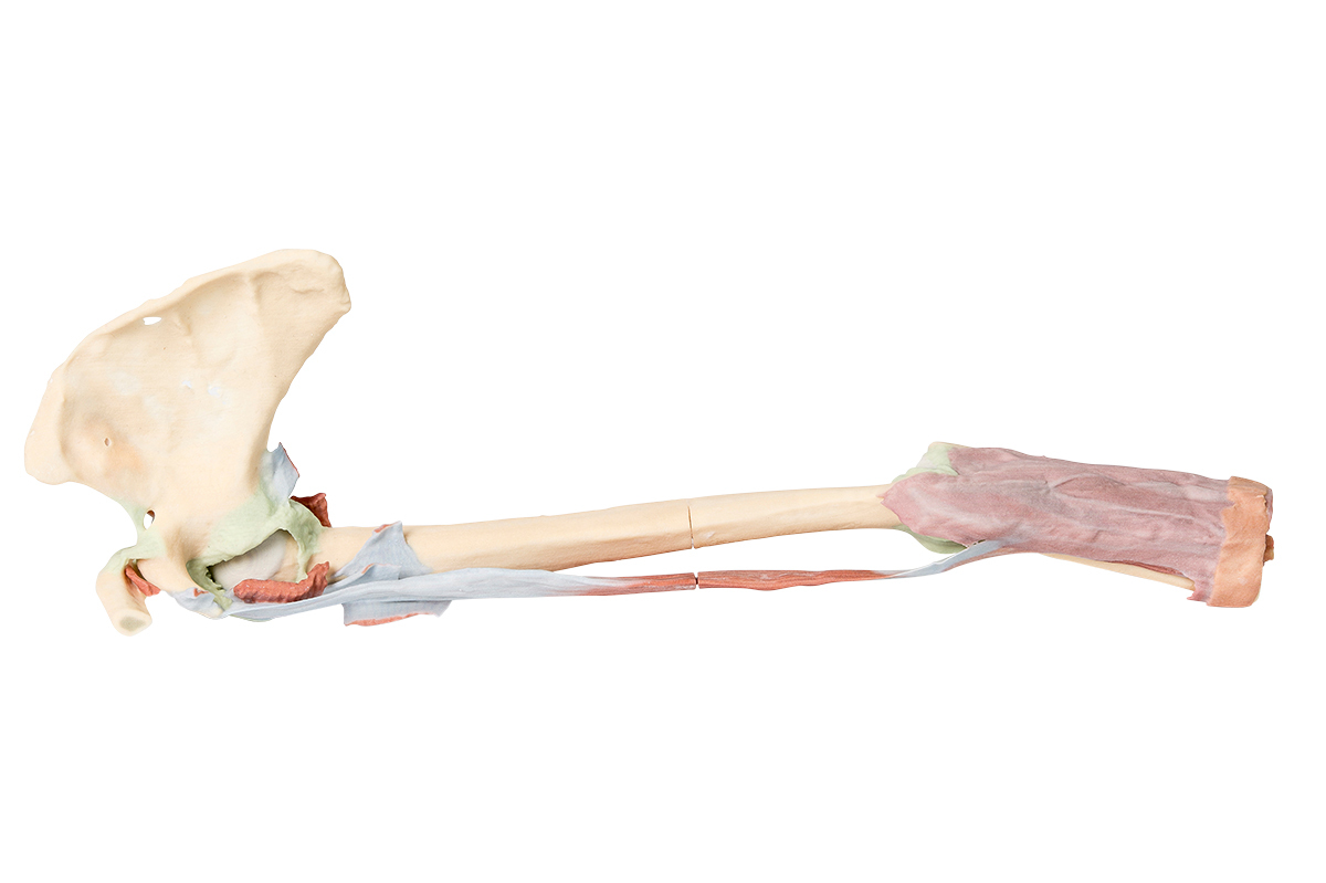
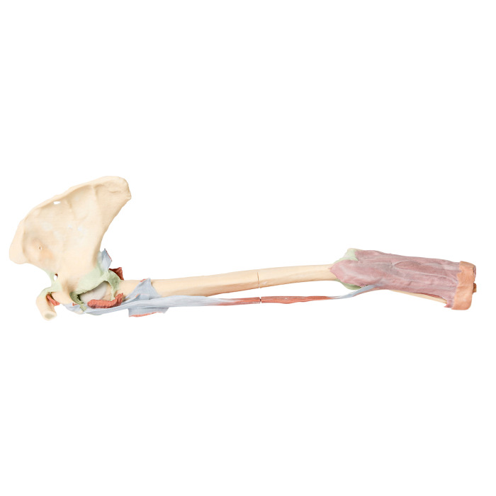
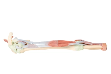
This 3D print shows the origin and insertion of biceps (most other arm and shoulder muscle bellies have been removed). The long head of biceps arises from the supraglenoid tubercle (hidden from view) and travels inferiorly in the bicipital groove, whereas the short head of biceps arises from the coracoid process. The bifid insertion of the muscle as the bicipital aponeurosis and the rounded tendon which can be seen winding around the radius to insert into the radial tuberosity are clearly discernable. At the shoulder region short stumps of some muscles (subclavius, subscapularis, pectoralis major, teres minor, infraspinatus, long head of triceps) and the tendinous insertion of latissimus dorsi can be identified close to the ‘floor’ of the medial lip of the bicipital groove. The tendon of teres major lies on the medial lip of the groove and the pectoralis major tendon inserts into the lateral lip of the groove. The tendon of pectoralis minor arises from the coracoid process medial to the origin of the short head of biceps. Ligaments of the shoulder region such as the coracoclavicular, coracoacromial, coracohumeral can be seen as can the capsule of the shoulder joint and that of the acromioclavicular joint. The supraspinatus muscle is the only rotator cuff muscle that has been completely preserved. The suprascapular ligament which bridges across the suprascapular notch is also evident on the superior border of the scapula.
At the elbow the capsule of the joint can be seen as can the annular ligament of the radius. The radial collateral ligaments are also just discernable. The ulnar collateral ligament is not visible as the two heads of flexor carpi ulnaris have been retained.
- Quantitative unit
- ks

MP1515 - Upper Limb - biceps, bones and ligaments


